


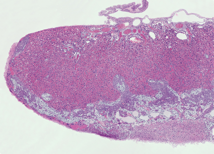
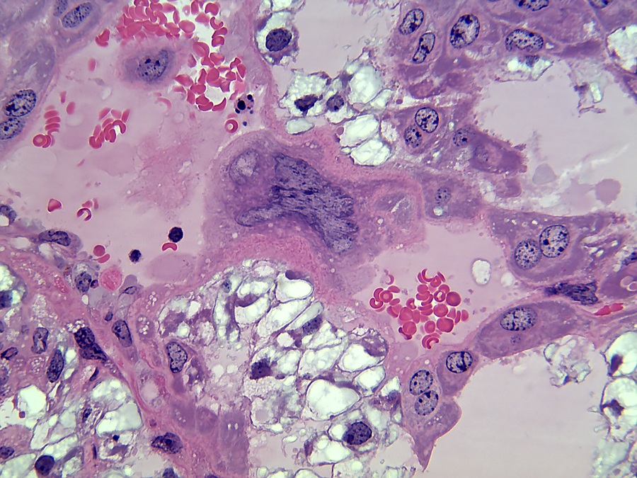
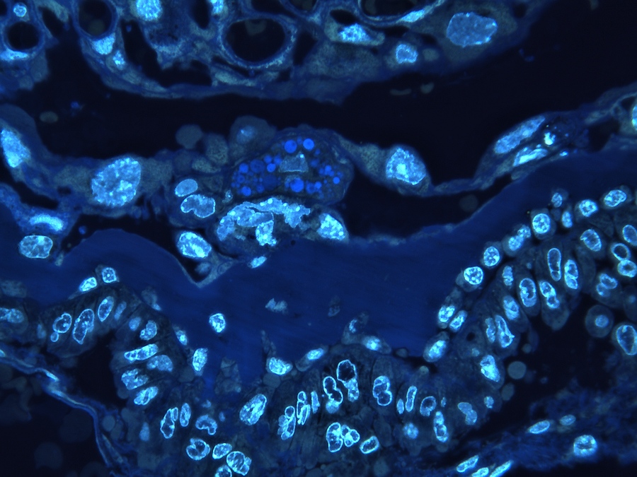
Trophoblast giant cells in the placenta of a mouse embryo.
Purpose of the Preparation: To embed a mouse placenta in Technovit 7100 and visualize the trophoblast giant cells.
Placenta[1]
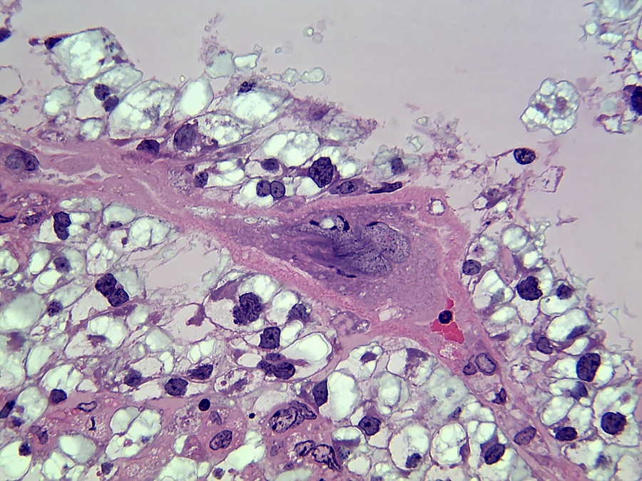
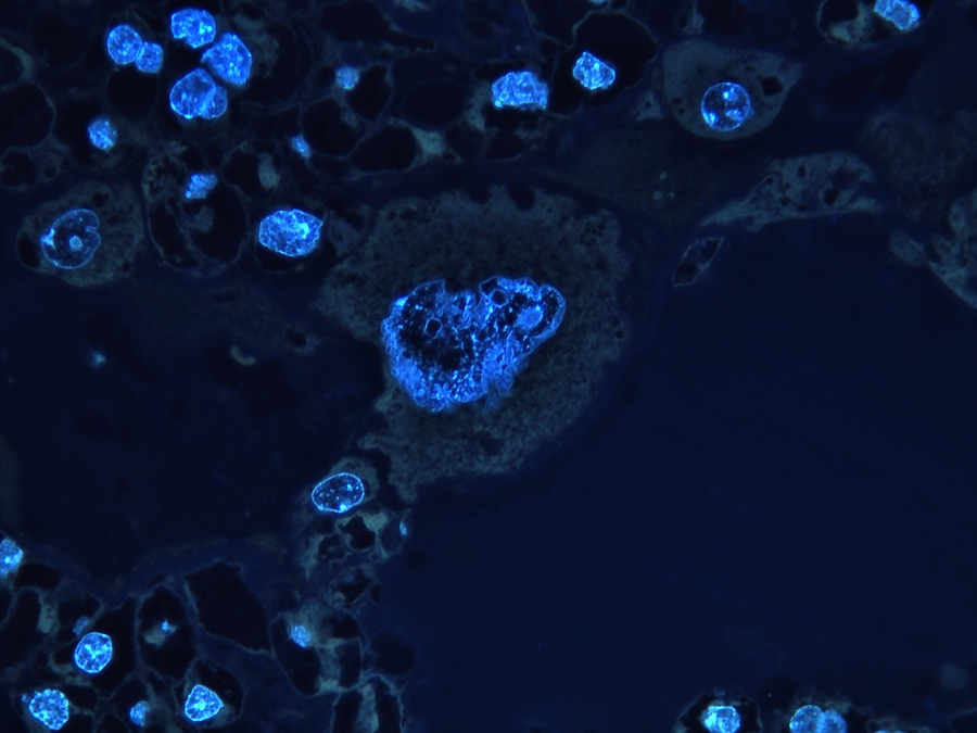
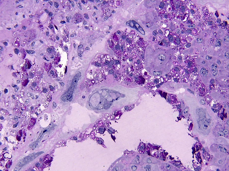
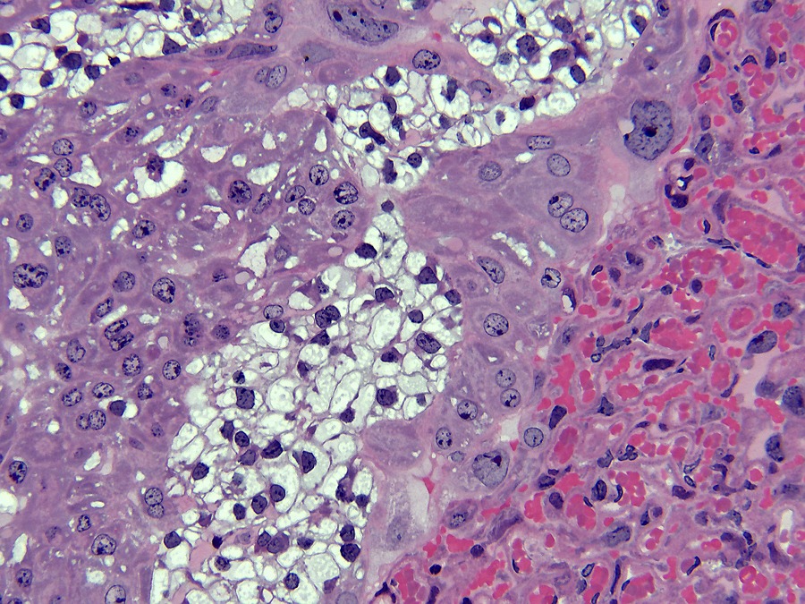
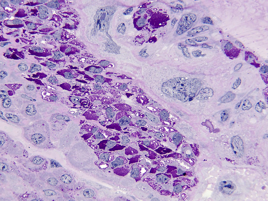
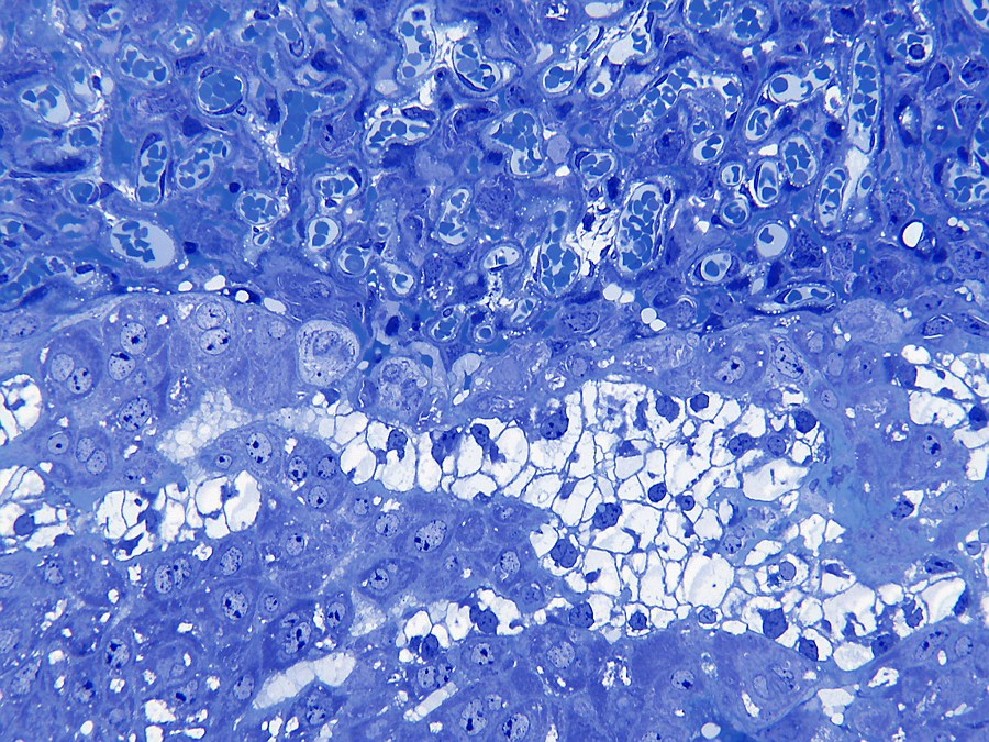
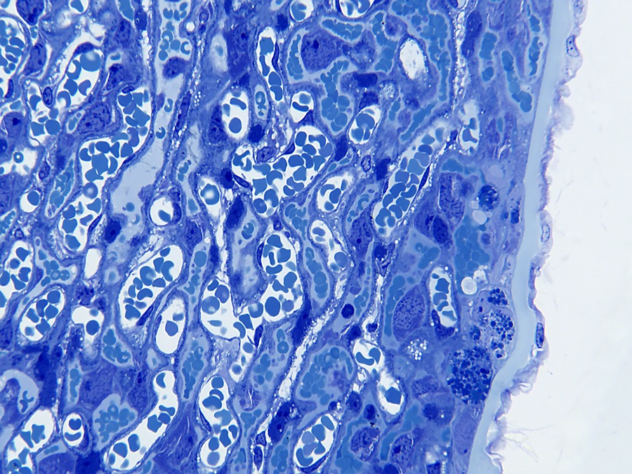
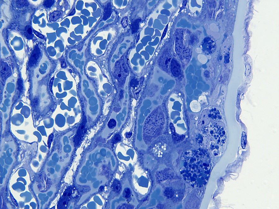
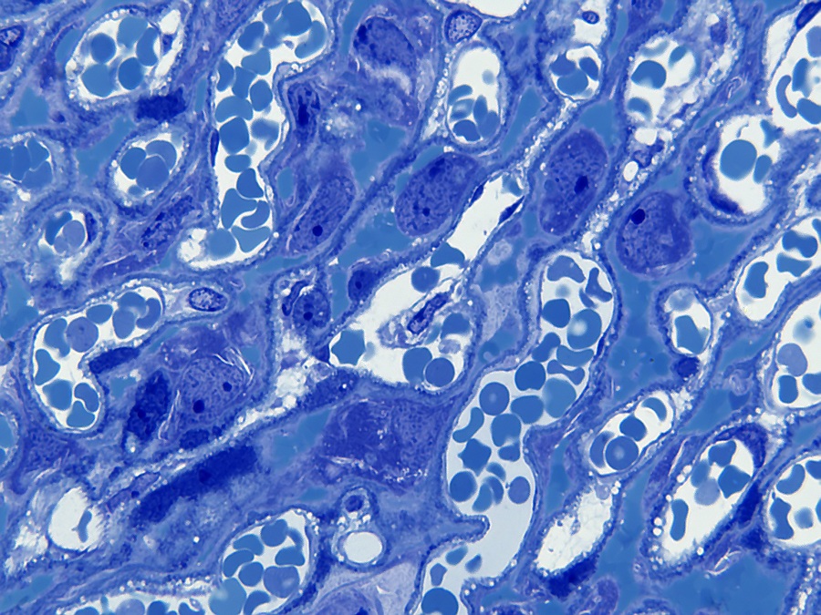
The mouse has a hemochorial placenta in which maternal blood is in contact with fetal chorion through a labyrinthine layer (humans have a similar type of placentation). The placenta consists of maternal and embryonic layers. The maternally derived layer is the decidua, which develops from the endometrium. The decidua consists of two parts: the decidua basalis and the decidua capsularis. The basalis has a rich vascular network and consists of loosely adherent irregular large vacuolate cells with differently shaped nuclei of different sizes. The decidua is separated from the embryonic labyrinthine zone by the junctional zone. The junctional zone contains maternal blood vessels (venous drainage), trophoblast cells and spongiotrophoblast cells. Trophoblast cells are generally very large with bizarrely shaped or multiple nuclei. Giant cell trophoblasts, as their name implies, are very large - up to 100 µm in diameter and are located closest to the decidua. Spongiotrophoblasts are phagocytic[6] and may contain phagocytized erythrocytes and are located closer to the labyrinthine zone. The labyrinthine zone consists of closely opposite fetal and maternal blood channels. The fetal blood channels are lined with thin endothelial cells and contain large immature nucleated red blood cells, while the maternal blood channels are lined with large labyrinthine trophoblast cells.
labyrinthine zone by the junctional zone. The junctional zone contains maternal blood vessels (venous drainage), trophoblast cells and spongiotrophoblast cells. Trophoblast cells are generally very large with bizarrely shaped or multiple nuclei. Giant cell trophoblasts, as their name implies, are very large - up to 100 µm in diameter and are located closest to the decidua. Spongiotrophoblasts are phagocytic[6] and may contain phagocytized erythrocytes and are located closer to the labyrinthine zone. The labyrinthine zone consists of closely opposite fetal and maternal blood channels. The fetal blood channels are lined with thin endothelial cells and contain large immature nucleated red blood cells, while the maternal blood channels are lined with large labyrinthine trophoblast cells.Giant cell trophoblast cells[3] are involved in the modulation of the maternal vasculature of the decidua and they produce several placental-specific hormones similar to the pituitary hormone prolactin[6]. These include Pl-1, Pl-2 (placentalactogen) and proliferin, which are secreted exclusively by giant cells. Pl-1 and Pl-2 modulate ovarian activity by stimulating the production of luteal progesterone, which is essential for the maintenance of pregnancy. In addition, Pl-1 and Pl-2 are involved in the control of mammary gland development.
[2] Stefanie L Bronson & Tracy L Bale, 07 August 2015, ‘The Placenta as a Mediator of Stress Effects on Neurodevelopmental Reprogramming.’, Fig. 1.

[3] Mark Kibschull, January 2003, ‘Trophoblast-Stammzellen als Modell zur Analyse der Conexin vermittelten Signalwege in der Plazentaentwicklung der Maus‘, Pag 2.
Materials and methods
One mouse was euthanized with ether. Embryos were removed at 15.5 dpc (Theiler stage 24)[4] and fixed in a mixture of formaldehyde 2%/glutaraldehyde 2.5%/phosphate buffer Sorensen Ph 7.17.
After dehydration to ethanol 100% was embedded in Kulzer's Technovit 7100[5]. Sections were cut on a LKB 2218 Historange microtome with a 'D' shaped hard steel blade.
Images were taken on a Leitz Orthoplan with Moticam 2300 camera.
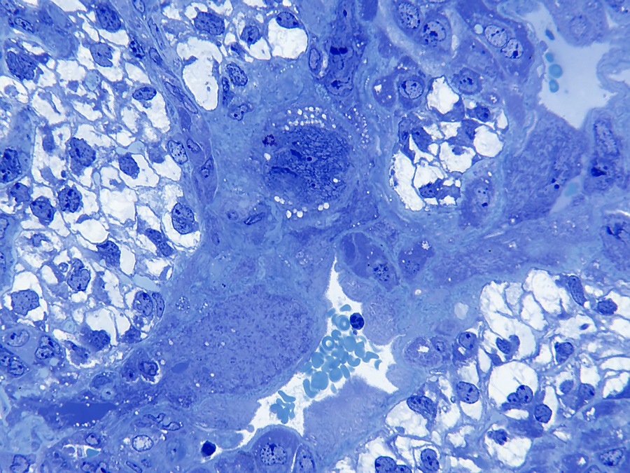
The zoomable image consists of 88 images with a Leitz PL 16x fluotar objective.
Overview image of placenta with zoomable image.
The image below gives an impression of the placenta. In the image to the right you can zoom in to the cellular level. The various layers can also be viewed.


Source reference:
[1] Cheryl L. Scudamore, A practical guide to the histology of the mouse, ISBN: 978-1-119-94120-0, Pag 96, 6.2.7 ‘Placenta’.
[2] Stefanie L Bronson & Tracy L Bale, 07 August 2015, ‘The Placenta as a Mediator of Stress Effects on Neurodevelopmental Reprogramming.’, Fig. 1.[3] Mark Kibschull, January 2003, ‘Trophoblast-Stammzellen als Modell zur Analyse der Conexin vermittelten Signalwege in der Plazentaentwicklung der Maus‘, Pag 2.
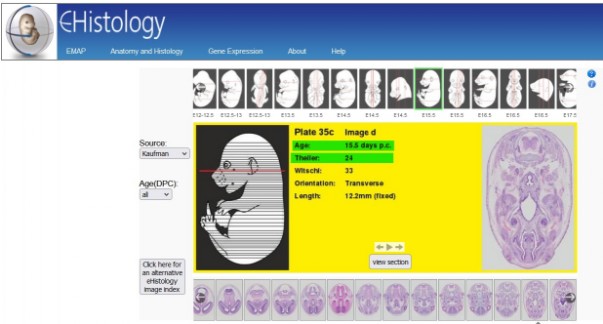
[4] Mouse foetus atlas: https://www.emouseatlas.org/emap/eHistology/kaufman/index.php?plate=38&image=38bb
[5] Technovit 7100 from the company Kulzer can be ordered from Morphisto in Germany. https://www.morphisto.de/en/products/technovitr-plastics/
[6] Wikipedia, https://en.wikipedia.org/wiki/Main_Page