


Update: 19-apr-2024
Welcome to the website.
The world under the miroscope shows how animal tissue is prepared into a microscopic slide. The histology includes the removal of tissue, fixation, embedding in paraffin or plastic. This is followed by cutting into coupes on a microtome and staining the specimen. Coupes are very thin slices of tissue. The slide is then examined under a microscope, photographed and the various cells are named with their theoretical underpinning. Attention is also focused on histological materials, microscopes, objectives and everything that has to do with a microscope. This small world is viewed with a Leitz Orthoplan transmitted light microscope equipped with high quality Fluotar and Apo objectives. All specimens shown are homemade and very diverse. Some examples of tissues and cells are: purkinje cells in the cerebrum, bacteria, cell division (mitosis and meiosis), blood cells, sperm, neurons, mucus cells, mast cells, kidney, pancreas with islets of Langerhans et cetera.
Some examples of specimen images.


Some examples of slides:
(the page 'Slides' is regularly expanded with new material)
Saggital section of a mouse, see sample description.
Because this coupe shows many different types of tissues in different relationships, a zoomable image was chosen. The section was photographed with a Moticam  2300 camera and here consists of 281 images. The images were stitched together with photoshop and a zoomable image was obtained with the program 'Zoomify'. Different cell types can be clicked in the legend.
2300 camera and here consists of 281 images. The images were stitched together with photoshop and a zoomable image was obtained with the program 'Zoomify'. Different cell types can be clicked in the legend.
Nissle substance in motor neurons, cresyl violet acetate staining
Preparation details,
The brain stem and cerebellum were taken from a Guinea pig and fixed in formaldehyde 4%.
The block was embedded in paraplast plus and then cut on an A&O 820 rotary microtome. Thickness is 4µm.
Depex from the company Serva was chosen as mount resin and can be purchased there, see sample discription for more details.


Sagittal coupe of an 8 day old mouse foot,
Click on the image to zoom in!
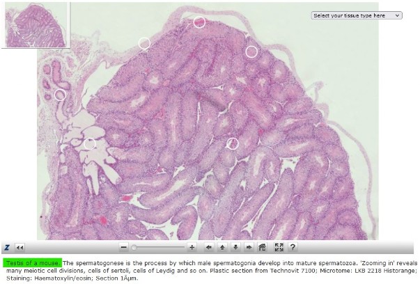
Spermatogenesis in the testis of a mouse,
Click on the image right to zoom in!
Lung vesicles (Alveoli) of a rat,
In the lungs (pulmones), which are located in the pleural cavity, the gas exchange takes place between the inhaled air and blood. The inhaled air enters the lungs via the bronchi (airways). The lungs consist of different structures, connected by connective tissue and covered by the pleura (lung membrane). The bronchi branch out at the top of the lungs to the left and right of the trachea. In the lung, the bronchus branches into secondary and tertiary bronchi and then further into bronchioli. The bronchioli branch out further into lung vesicles, the alveoli.
Look at the slide page for more theory and beautiful images.

Cutting Technovit 7100 on an LKB 2218 Historange microtome,
This type of microtome with retraction was developed specifically for cutting plastics and was produced by the company LKB bromma in Sweden. The year of production of the Historange is 1982. See: http://www.lkbprod.com/
Two documents about the Historange can be downloaded from the download page. The "user manual" and the "part breakdown.
The microtome is, by the author, only used for plastics and mainly Technovit 7100 is cut with it.


Sagittal coupe of an unborn mouse 15.5dpc,
In mouse embryology, 15.5dpc (15.5 days post coitum = 15.5 days after insemination) is also called 'Theiler stage' 24. This sample is fixed with Bouin, molded in paraffin and cut 2 µm thick. The coupe is stained with Haematoxylin/eosin (HE staining). Click on the image to view the coupe. Many organs are already clearly visible such as heart, brain, lungs and liver. Look at the Slides page for more information.

Epon epoxy cutting on an ultramicrotome,
On this Reichert-Jung Ultracut ultramicrotome a coupe is cut out of Epon.
Take a look at the 'Microtomes' page to read more information about this type of microtome.
Contact: microscoop@ronaldschulte.nl
Disclaimer: All images of slides, equipment and videos shown on this website have been produced by the author and are therefore not for free use. You must ask permission for publication in any form at all times.